About 20 years ago, a neighbor knocked on my door and very sheepishly asked me if I would answer a question for her. She knew I worked in the medical field and that’s why she was at my door. She told me that her doctor had just advised her that she needed an MRI and that he would see her back after it was performed. She very quietly said to me “I’ve never had one before. What happens with an MRI? Does it hurt? Do I have to take my clothes off? Do I have to fast before I have it?”. She didn’t feel safe asking these questions of her doctor, which is unfortunate, but common.
Let’s talk about common diagnostic tests that you might find yourself having to undergo. Like my neighbor who didn’t know anything about an MRI, there is often confusion surrounding these studies and sometimes, ignorance about the risks can be harmful to your health. I’ll explain that in a bit. Let’s define some things first.
We are exposed to natural sources of radiation all the time. According to recent estimates, the average person in the U.S. receives an effective dose of about 3 mSv (millisieverts) per year of natural radiation, which includes cosmic radiation from outer space. (A millisievert is a standard unit of measurement of x-ray dosage). These natural “background doses” vary according to where you live. People living at high altitudes such as Colorado receive about 1.5 mSv more per year than those living near sea level. A coast-to-coast round-trip airline flight is about 0.03 mSv due to exposure to cosmic rays. Interestingly, the largest source of background radiation comes from radon gas in the ground underneath our homes (about 2 mSv per year). Like other sources of background radiation, the amount of radon exposure varies widely depending on where you live.
Radiation is not stored in the body, but the effects of a person being exposed to radiation add up over time. With each exposure to radiation a person has in their lifetime, there is increased risk of harm. A small dose of radiation (like one dental x-ray) carries very low risk. The higher the dose of radiation, the greater the risk.
So what about x-rays? In standard X-rays, a beam of energy is aimed at the body part being studied. Its quick and easy and is really just a picture of the inside of the body. How hazardous are they? To put it simply, the amount of radiation from one adult chest x-ray (0.1 mSv) is about the same as 10 days of natural background radiation that we are all exposed to as part of our daily living.
CT stands for ‘computed tomography’. Many years ago, it used to be referred to as “CAT scan”, for computer-assisted tomography, but that term has become obsolete. CT scans are more detailed than standard x-rays. In CT, the x-ray beam moves in a circle around the body. This allows many different views of the same organ or structure and provides much greater detail. The x-ray information is sent to a computer that interprets the x-ray data and displays it in two-dimensional form on a monitor. Sometimes CT scans are done with contrast, which is infused via an IV line. This contrast lights up the blood vessels and allows better visualization of certain structures. If you have a CT scan of the abdomen, you may be asked to drink an oral contrast, which then lights up your digestive tract for better visualization. CT scanning is not painful and is fairly quick. You lay on a table that moves you into a large donut-shaped machine and while inside, you may hear some clicking noises but these are not as loud as an MRI machine. However, CT scans come with a lot of radiation. One CT scan equals a lot of x-rays. As an example, a CT of the chest is comparable to about 2 years of natural background radiation. A CT scan of the brain without contrast is comparable to about 7 months of natural background radiation. The effects of radiation are cumulative, as I stated before. Here’s an example of what hopefully doesn’t happen.
I recently reviewed the medical records for a legal case that involved a young woman who was 26. She got ill at age 20 and between the ages of 20-22, she had 12 CT scans of her abdomen and pelvis. Twelve. And no doctor ever caught the fact that she was having these serial CT scans. They were mostly ordered by different ER physicians who didn’t know much of her history, so they just kept ordering them. The young woman didn’t say anything because she knew nothing about the radiation risks inherent in CT scans. That woman may have already reached her lifetime limit of acceptable radiation, and she’s only 26.
Now, don’t get me wrong, CT scans can be lifesavers and are a wonderful diagnostic tool. But if your doctors are not aware that you have already had many CT scans in the last few years, it’s really proactive on your part to inform them of this so that an alternative method might be considered.
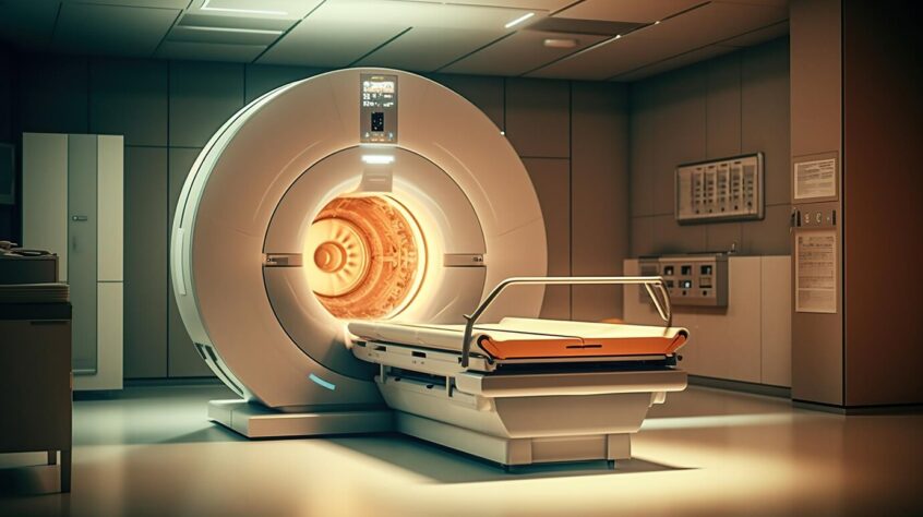
That brings me to MRI. Magnetic Resonance Imaging. No radiation. None. An MRI unit is a gigantic magnet. Because the MRI unit is a giant magnet, no metal is allowed in the MRI room. That means you cannot wear anything metal on your body and if you have any type of shrapnel or bullets or wire mesh in your body, you can’t have an MRI. In 2001, a 6-yr-old boy was killed inside of an MRI unit when a trainee came into the MRI room with an oxygen tank. The magnet turned the tank into a torpedo which was drawn into the unit and killed the boy. Recently, a woman was in an MRI unit in Northern CA and someone brought a non-MRI (metallic) wheelchair into the room where she was and it was sucked into the unit. The woman was miraculously unharmed. These are exceedingly rare situations, but they have happened.
An MRI can also be done with contrast through an IV line. It takes longer than a CT scan, typically 20-30 minutes, and you must lie very still for that entire time. It you are claustrophobic, you may need anxiety medication beforehand because it is a very tight and loud environment. However, MRIs are an extremely good diagnostic tool and can show things that cannot be seen well with other modalities. If you have a torn ACL in your knee, for instance, this won’t show on an x-ray, but it will show up on MRI.
Another commonly used diagnostic tool is ultrasound. Ultrasound uses sound waves, does not involve radiation, and can be used well in many instances.
Fluoroscopy is a medical procedure that makes a real-time video of the movements inside a part of the body. Images are captured by passing x-rays through the body over a period of time. Radiation doses are usually higher than in regular x-rays. Fluoroscopy is used in barium enemas and esophagus swallowing studies as well as guidance in many procedures, including angiograms.
The diagnostic tool with the highest radiation is a PET scan – positron emission tomography. It is used in conjunction with CT and is often called a PET/CT scan. It is done over the whole body and is typically used to determine if a cancer has spread to other parts of the body. One PET scan is comparable to 7.6 years of natural background radiation.
This is not a comprehensive list of diagnostic modalities in use today, and my intention is definitely not to talk you out of getting any of these tests. On the contrary. These are wonderful diagnostic tools. As a patient advocate, my desire is to raise your awareness with the goal of helping you become a more proactive patient. The best advice is to speak with your physician about your concerns, get your questions answered, get only the imaging tests that are needed, and try to limit your exposure to all forms of radiation. Good communication is always key in achieving this goal.

